
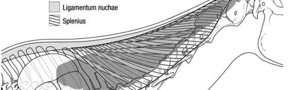
Variations of the Nuchal Ligament Lamellae
One of the functions of the Nuchal ligament is to passively support the head and neck while countering against gravity. This study investigated the anatomy and insertion of the Nuchal ligament lamellae to the cervical vertebrae (C2 to C7). Furthermore, it explains the biomechanical relevance of these findings.
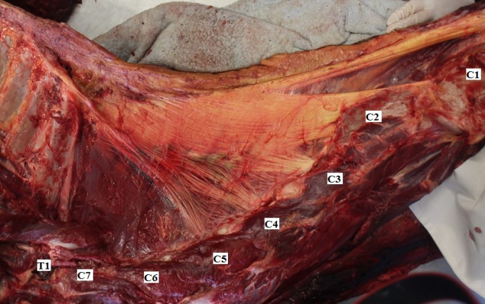
The cervical region of 35 horses (mixed gender and breed, aged between 2 and 28 years) was dissected until the anatomy of the Nuchal ligament lamellae was revealed. The lamellae of all horses showed insertion points from C2 to C5 (see image below). Whereas no single one horse showed insertion of the lamellae to C2–C6 or C2-C7.
Earlier research showed that the largest angular difference in cervical flexion occurs at C1 and C6 and the largest lateral bending at C6. However, another kinematic study found that lateral bending was greatest at the cervico- thoracic junction (C7–T1) and that axial rotation was the second and third highest at C6–C7 and C7–T1, after C1-C2.
This article suggests that increased motion in the caudal cervical spine might be the result of the absence of the lamellae on C6 and C7. Knowing the anatomy of the Nuchal ligament lamella, may lead to a further understanding of the pathogenesis of facet joint arthropathy in the caudal cervical vertebrae. This radiographic study gives further information about caudal cervical facet joint arthropathy
> From: May-Davis et al., Equine Vet J. 34 (2017) 1110-1113. All rights reserved to Elsevier Inc.. Click here for the online summary.